Scintigraphy
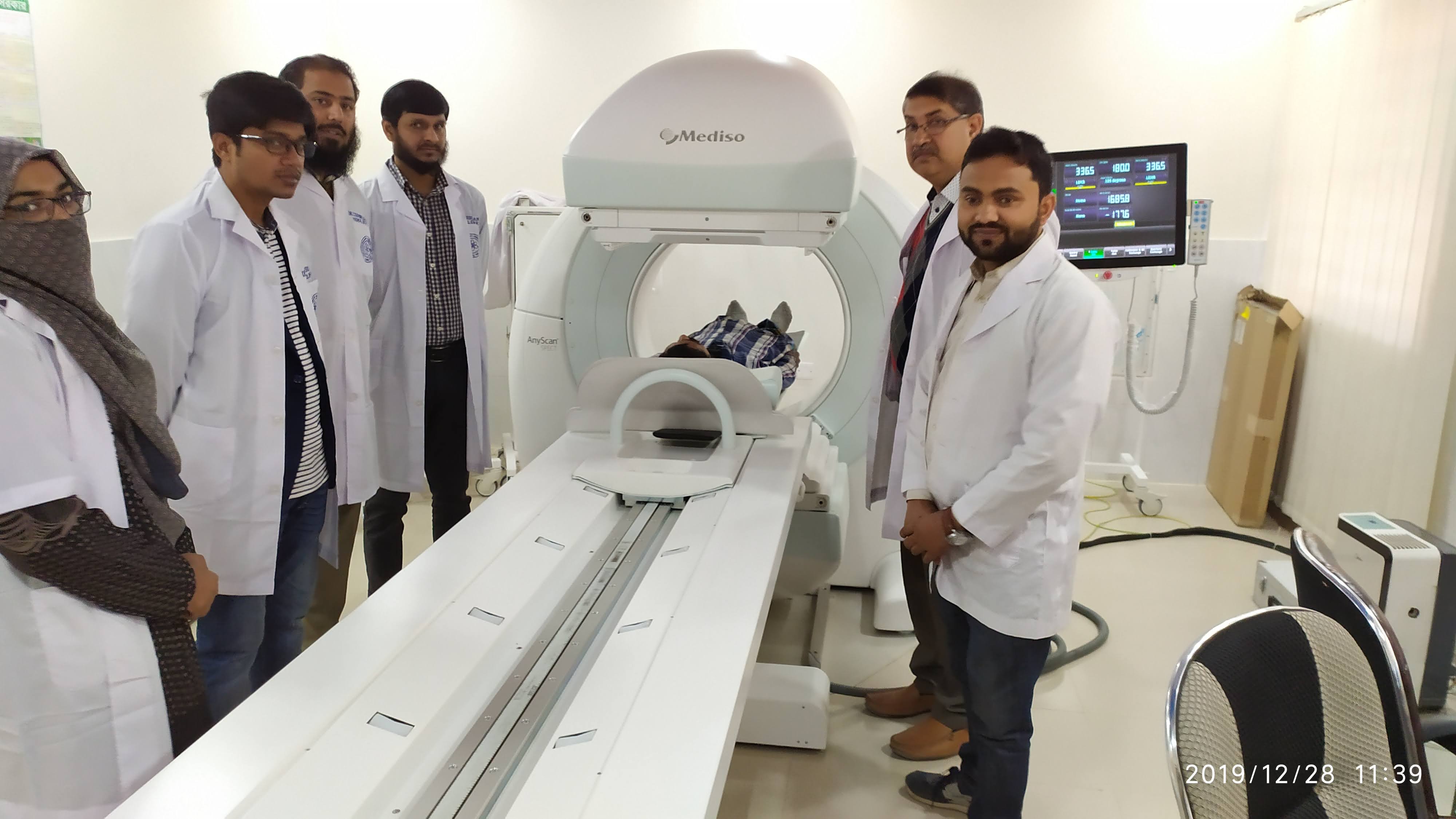
The special camera and imaging techniques used in nuclear medicine include the gamma camera and single-photon emission computed tomography (SPECT). The gamma camera, also called a scintillation camera, detects radioactive energy that is emitted from the patient's body and converts it into an image. It simultaneously detects radiation from the entire field of view and enables the acquisition of dynamic as well as static images of the area of interest in the human body.
Nuclear medicine imaging uses small amounts of radioactive material to diagnose, evaluate or treat a variety of diseases. These include many types of cancers, heart disease, gastrointestinal, endocrine or neurological disorders and other abnormalities. Because nuclear medicine exams can pinpoint molecular activity, they have the potential to identify disease in its earliest stages. They can also show whether a patient is responding to treatment. Nuclear medicine imaging procedures are non-invasive. With the exception of intravenous injections, they are usually painless.
Working Schedule
Working hour
Every Saturday to Thusday at 8.00am -2.30pm
Except Friday and all government holiday
Service Start Time
Every Saturday to Thusday at 7.30 AM
Except Friday and all government holiday
The department started the service at 7.30 am
(The certain patient will be appointed according to the availability of Radio Isotopes and Kit)
Alert
| Investigations | Rate | Preparation |
|---|---|---|
| DMSA-Renal Scan | 800 | Get Appointment |
| DTPA-Brain Scan (Tc-99m) | 800 | Get Appointment |
| DTPA-Renogram | 1000 | Get Appointment |
| Hepatobiliary scan (Tc-99m) | 1200 | Get Appointment |
| Hysterosalphingo Scintigraphy (Tc-99m) | 1000 | Get Appointment |
| I-131 Thyroid Scan | 500 | Get Appointment |
| Liver Spleen Scan (Tc-99m) | 600 | Get Appointment |
| Meckels Diverticulums Scan | 1000 | Get Appointment |
| Three phase Bone scan (Tc-99m) | 1500 | Get Appointment |
| Thyroid Scan (Tc-99m) | 500 | Get Appointment |
| Thyroid uptake study | 400 | Get Appointment |
| SPECT Bone Scan | 2500 | Get Appointment |
| SPECT Kidney Scan | 1200 | Get Appointment |
| SPECT Liver Scan | 1200 | Get Appointment |
| SPECT CT WHOLE BODY SCAN | 3000 | Get Appointment |
Depending on the type of exam, the radiotracer is injected, swallowed or inhaled as a gas. It eventually accumulates in the area of the body under examination. A special camera or imaging device detects radioactive emissions from the radiotracer. The camera or device produces pictures and provides molecular information.
Many centers superimpose nuclear medicine images with computed tomography (CT) or magnetic resonance imaging (MRI) to produce special views. This is known as image fusion or co-registration. These views allow the doctor to correlate and interpret information from two different exams on one image which leads to more precise information and accurate diagnoses. Single photon emission computed tomography/computed tomography (SPECT/CT) and positron emission tomography/computed tomography (PET/CT) units can perform both exams at the same time.
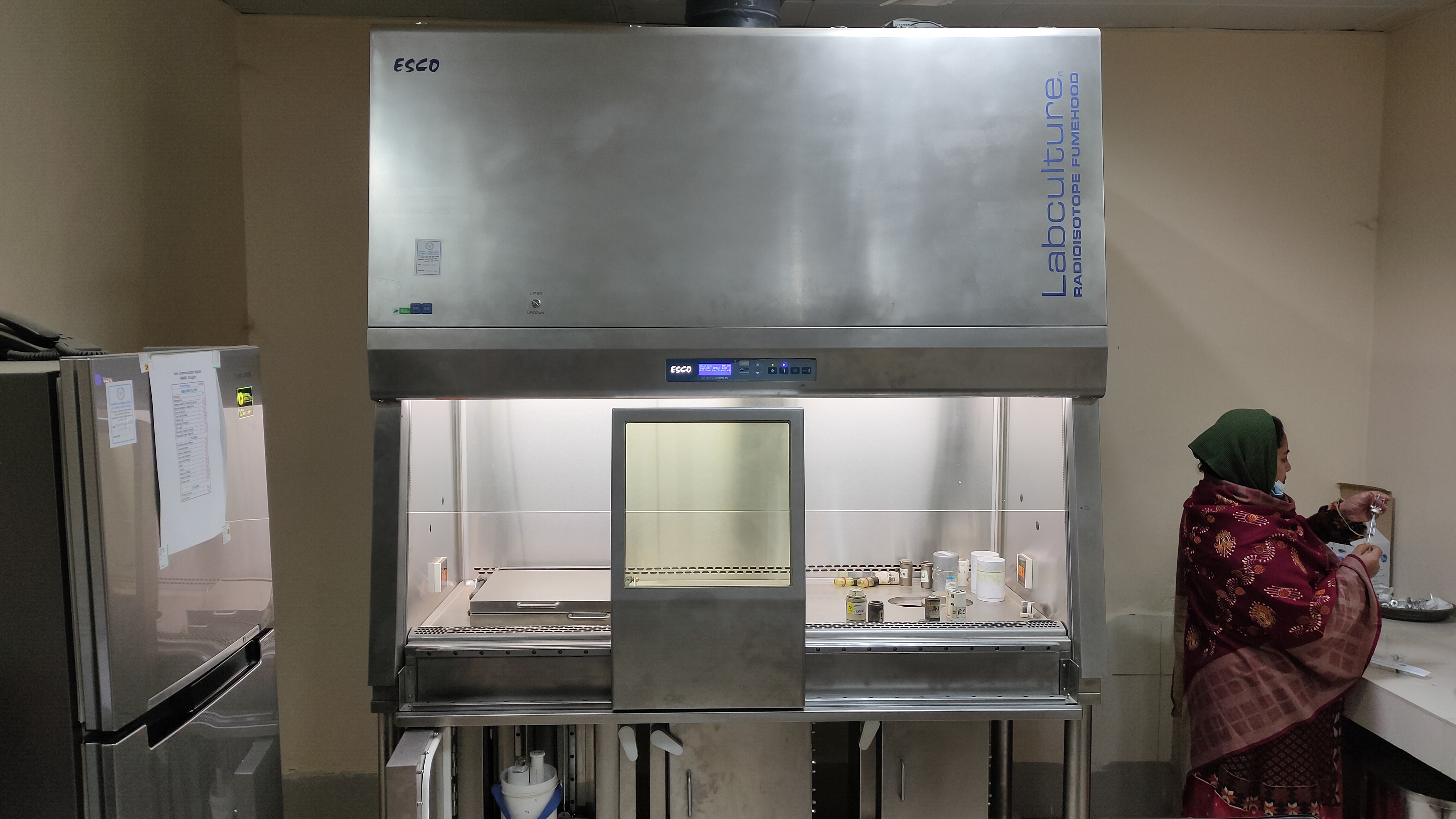
Following scintigraphy services are provided by the INMAS, Sylhet:
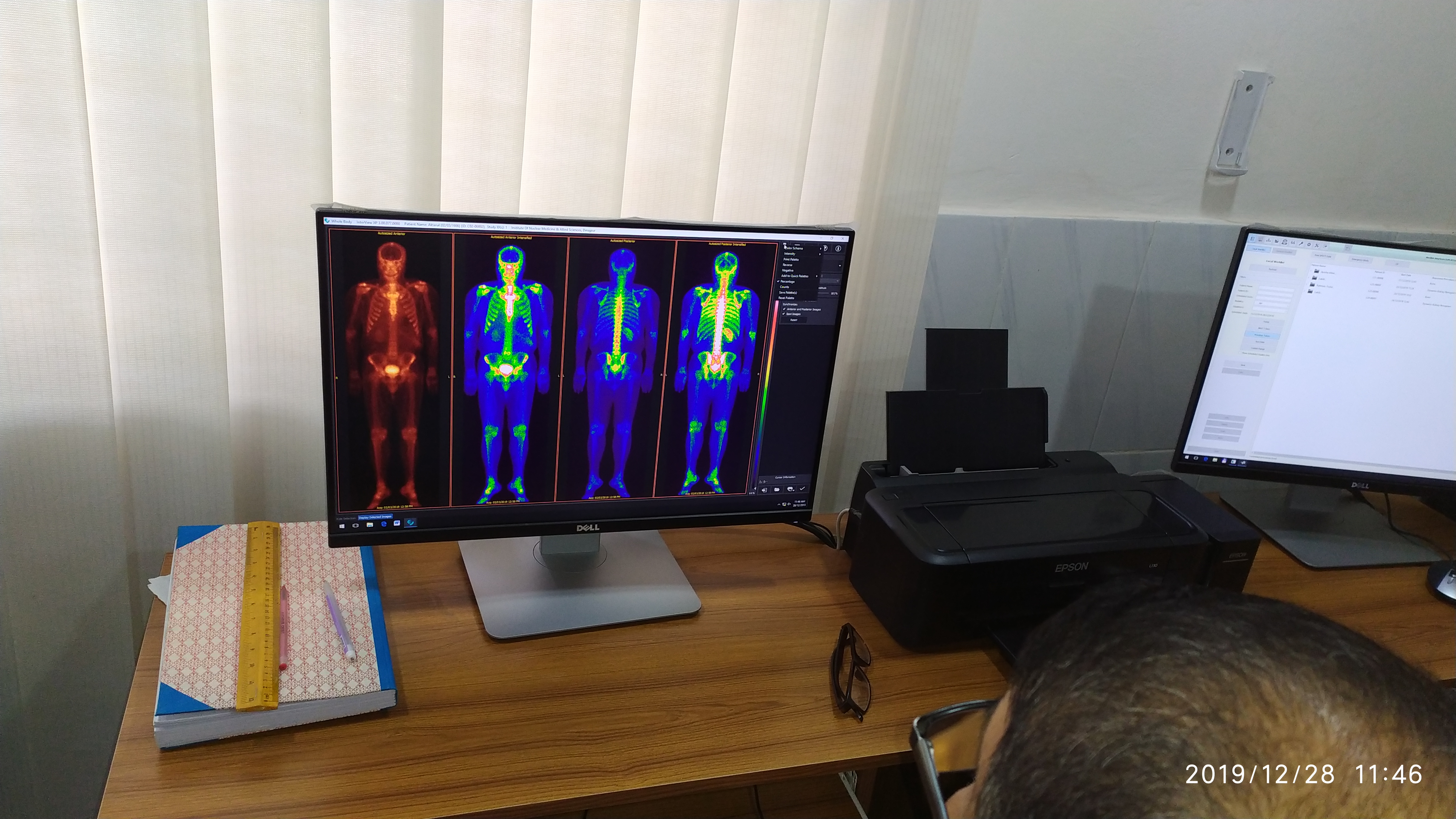
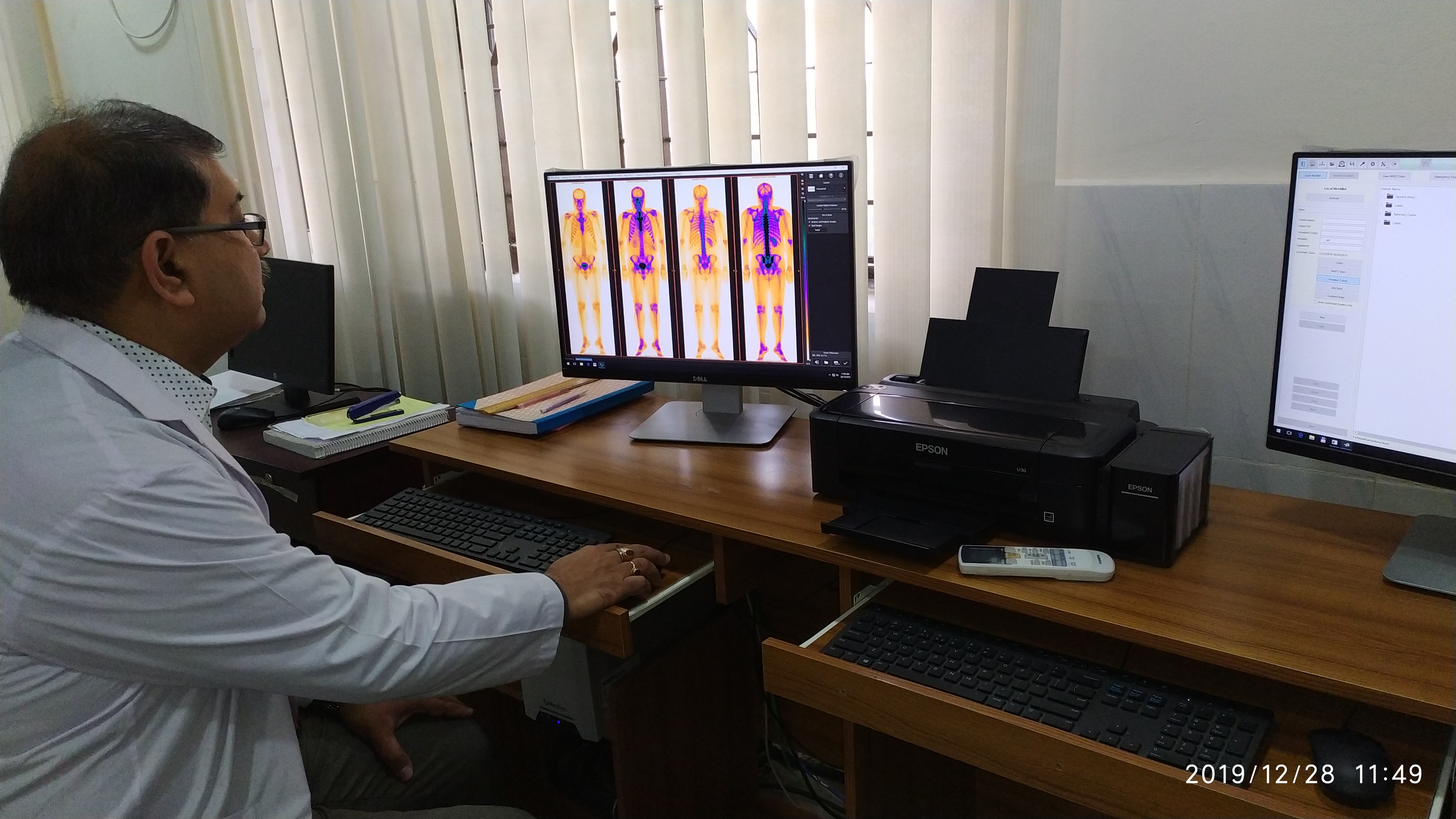
Imaging of Skeletal System
Done by 99mTc MDP (methyl diphosphonate)
Done for –
1. Detection of metastasis or follow up
2. Differentiation between osteomyelitis and cellulitis
3. Bone viability or avascular necrosis detection
4. Stress fracture, prosthesis evaluation.
Renal Scintigraphy
The available tests are:
1. DTPA-Renogram and Split renal function:
Done by- 99mTc-DTPA (diethylene-triamine-pentaacetate)
Helps in distinguishing between obstructive hydronephrosis and non-obstructive collecting system dilatation of the kidneys.
Evaluation of medical and surgical complications of renal transplant e.g. acute tubular necrosis, rejection and surgical mishaps like urinoma, lymphocele, hematoma, ureteral obstruction and vascular complications.
2. DTPA-Renogram with camera GFR(Tc-99m): GFR can be detected early and more accurately before any change in serum creatinine level.
3. DMSA-Renal scan (Tc-99m):
Done by- 99mTc-DMSA (dimercaptosuccinic acid)
Detection of structural and congenital abnormalities, tumor and other causes of metastases in some cases.
Thyroid Scintigraphy
1.Thyroid uptake test:
Done by- 131-I
Used to see the functional status of thyroid gland.
2.Thyroid scan:
Done by- 99mTc pertechnetate.
Used to see the size and shape of thyroid, congenital thyroid abnormalities and evaluate the amount of residual thyroid tissue after surgery.