
Ultrasound & CD
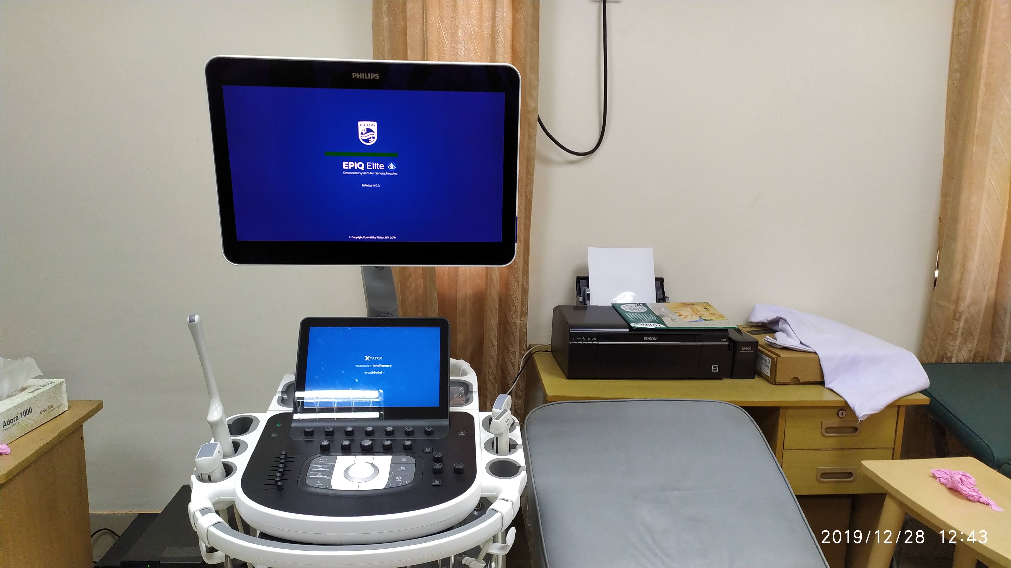
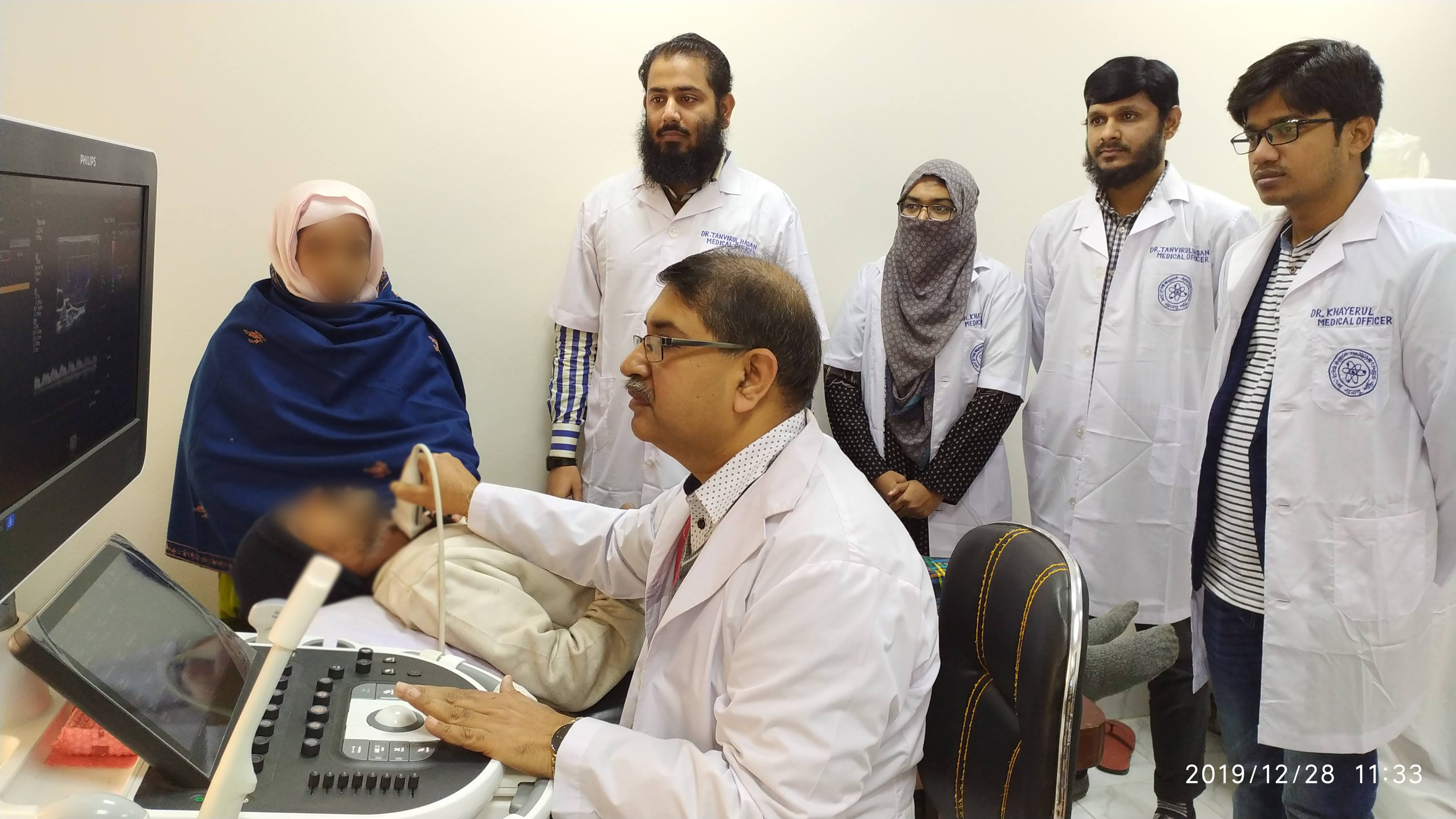
Ultrasound (also known as diagnostic sonography or ultrasonography): is a type of imaging. uses high-frequency sound waves to look at organs and structures inside the body. Doctors use it to view the heart, blood vessels, kidneys, liver, and other organs. During pregnancy, doctors use ultrasound to view the fetus. Unlike X-rays, ultrasound does not expose you to radiation.
Ultrasound is safe and painless. It produces pictures of the inside of the body using sound waves. Ultrasound imaging is also called ultrasound scanning or sonography. It uses a small probe called a transducer and gel placed directly on the skin. High-frequency sound waves travel from the probe through the gel into the body. The probe collects the sounds that bounce back. A computer uses those sound waves to create an image.
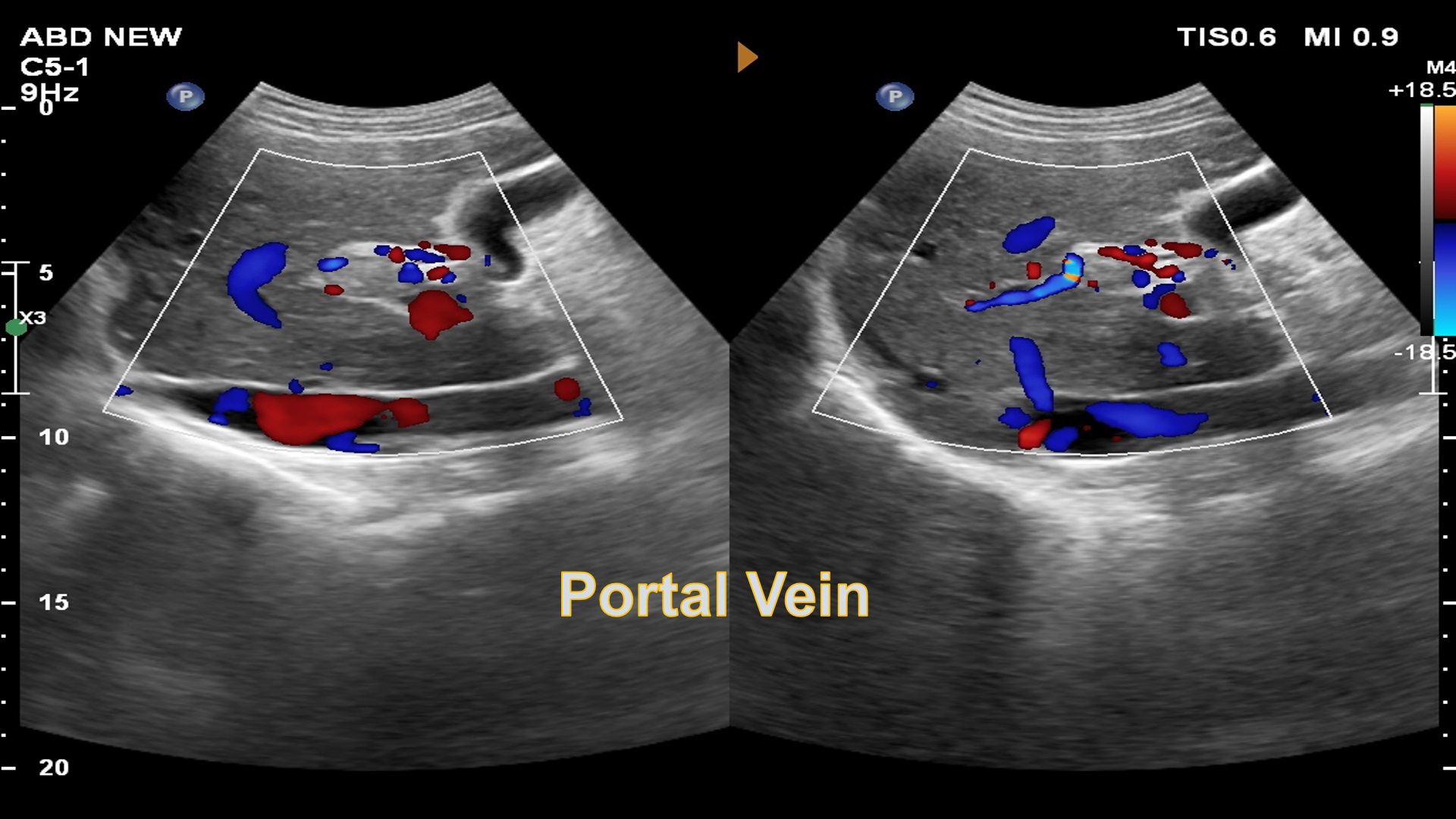
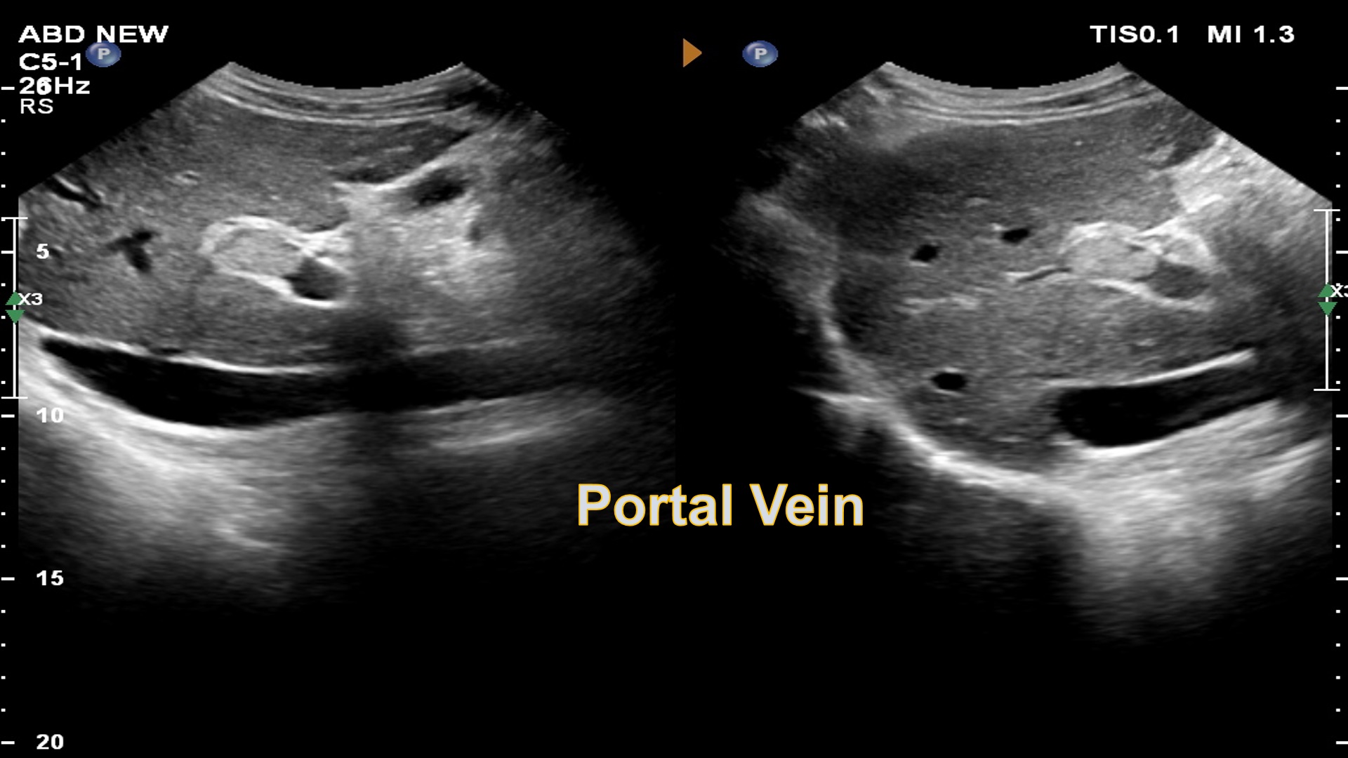
Images are captured in real-time, they can show the structure and movement of the body's internal organs. They can also show blood flowing through blood vessels.
Ultrasound imaging is a non-invasive medical test that helps physicians diagnose and treat medical conditions.
The use of ultrasound in medicine began during and shortly after the 2nd World War in various centres around the world. The work of Dr.Karl Theodore Dussik in Austria in 1942 on transmission ultrasound investigation of the brain provides the first published work on medical ultrasonics. The application of ultrasound in medicine began in fifties of last century. First was introduced in the obstetrics, and after that in all the fields of the medicine (the general abdominal diagnostics. the diagnostics in the field of the pelvis, cardiology, ophthalmology and orthopedics and so on).
Conventional ultrasound displays the images in thin. flat sections of the body. Advancements in ultrasound technology include three-dimensional (3-D) ultrasound that formats the sound wave data into 3-D images.
Working Schedule
Working hour
Every Saturday to Thusday at 8.00am -2.30pm
Except Friday and all government holiday
Service Start Time
Every Saturday to Thusday at 7.30 AM
Except Friday and all government holiday
Alert
| Investigations | Rate | Preparation |
|---|---|---|
| USG of KUB | 300 | Get Appointment |
| USG of KUB+PVR | 400 | Get Appointment |
| USG of Lower abdomen | 300 | Get Appointment |
| USG of Pregnancy Profile | 300 | Get Appointment |
| USG of HBS/Upper abdomen | 300 | Get Appointment |
| HRUS of breast | 400 | Get Appointment |
| HRUS of breast & axilla | 500 | Get Appointment |
| Transvaginal Sonography | 700 | Get Appointment |
| HRUS of Local Part (Chest, Neck, Superficial organ etc.) | 400 | Get Appointment |
| HRUS of scrotum | 400 | Get Appointment |
| HRUS of thyroid | 300 | Get Appointment |
| Duplex Evaluation of Abdominal Tumor | 1000 | Get Appointment |
| Duplex Evaluation of Carotid & Vertebral Arteries | 1000 | Get Appointment |
| Duplex Evaluation of Uterus & Adnexa | 800 | Get Appointment |
| Endocavitary color Doppler (TVS/TRUS) | 1200 | Get Appointment |
| Obstetric Duplex (Pregnency, Fetal velocimetry/Fetal Echo) | 1000 | Get Appointment |
| USG of Whole Abdomen | 300 | Get Appointment |
| Fetal Anomaly Scan | 1000 | Get Appointment |
A Doppler ultrasound study may be part of an ultrasound examination. Doppler ultrasound is a special ultrasound technique that evaluates movement of materials in the body. It allows the doctor to see and evaluate blood flow through arteries and veins in the body.
Most ultrasound exams are painless, fast and easily tolerated. After an ultrasound examination, you should be able to resume your normal activities immediately. Standard diagnostic ultrasound has no known harmful effects on humans.